Frozen Shoulder Not Improving? It Might Actually Be Calcific Tendinitis! Full Analysis of Symptoms, Causes, and Treatments

Fang-Wei Hsu (Orthopedic Physician, Regen Clinic)
2025-05-15
What Is Calcific Tendinitis? Can Shoulder Tendons Develop Calcium Deposits?
Calcific Tendinitis is a condition where calcium deposits form within the shoulder tendons, most commonly affecting the rotator cuff tendons, especially the supraspinatus tendon. These calcium accumulations can trigger localized inflammation and pain, and in severe cases, may impair shoulder movement, daily activities, and sleep quality.
The main cause of calcific tendinitis is related to tendon injury. During the body’s self-repair process, cartilage-like cells may form in the tendon. If healing is incomplete, calcium-like deposits develop near the tendon, leading to poor tendon repair and inflammation.
Calcific Tendinitis Can Be Classified into Two Main Types:
Reactive Calcification: Caused by repeated injuries, overuse, recurrent inflammation, immune issues, patient constitution, metabolic abnormalities, or endocrine disorders.
Degenerative Calcification: Caused by long-term wear and aging of the shoulder rotator cuff tendons, poor tendon blood supply, or shoulder joint bone spurs.
Common Symptoms:
1. Severe shoulder pain (painful even at rest)
2. Limited active movement (difficulty raising the arm)
Symptoms According to Calcification Stage:
- Pre-calcification Stage: No noticeable symptoms in the early phase
- Calcification Formation Stage: Increased pain, most painful during arm abduction
- Calcification Resorption Stage: Intense pain, difficulty sleeping at night
- Calcification Maturation Stage: Calcification subsides, pain decreases
Although calcific tendinitis can occur at any age, certain groups are particularly prone to it or should be especially vigilant:
- Adults aged 30–50: Most patients are middle-aged adults, and the condition is associated with long-term use of the shoulder joint or tendon aging.
- Individuals who use their shoulders frequently: such as athletes (swimmers, tennis players, badminton players), laborers, painters, and those who spend long hours using computers or typing. Prolonged arm elevation or repetitive shoulder movements increase the strain on the tendons.
- Individuals with metabolic or endocrine disorders: For example, those with diabetes, thyroid dysfunction, or dyslipidemia, as these conditions can impair tendon repair and promote calcium deposit formation.
- Individuals with a history of shoulder injuries or chronic shoulder pain: Tendons that have already undergone chronic wear and tear or inflammation are more prone to calcification.
- Women: Statistics show that women are slightly more affected than men, which may be related to physical constitution or hormones.
In summary, individuals who engage in repetitive shoulder use, high-intensity exercise, or those with chronic tendon damage or metabolic disorders are the groups that need to pay the most attention to shoulder health and take steps to prevent calcific tendinitis.
- Women aged 30-50
- Throwing athletes (e.g., baseball players)
- Regularly lifting heavy objects
- Frequently raising arms (e.g., writing on boards, painters)
- Jobs requiring frequent keyboard use
- History of shoulder injuries
Calcific Tendinitis VS. Frozen Shoulder — Confused About the Difference?
The symptoms of calcific tendinitis and frozen shoulder are very similar, including common age range, night pain, limited mobility, and pain during movement. Since frozen shoulder is caused by joint adhesion and stiffness, it is usually less painful at rest, whereas calcific tendinitis can still cause pain even when not moving.
| Condition | Calcific Tendinitis | Frozen Shoulder |
| Age | 20-40 years old | 40-60 years old |
| Cause | Friction from calcium crystal deposition | Shoulder joint capsule contracture |
| Distinguishing Features | Sudden acute severe pain | Pain during movement |
| Pain occurs at certain angles of movement | Limited mobility at multiple angles | |
| Someone else can help lift the shoulder | Even with help, shoulder cannot be fully raised |
How is Calcific Tendinitis Diagnosed? What Examinations Are Needed?
1) X-ray – Clearly shows white calcifications, resembling stones; the most common initial examination.
2) Ultrasound – Can locate the calcification, assess tendon thickness, check for tears, and evaluate inflammation (can also be used for ultrasound-guided calcification aspiration and injection treatment).
3) MRI – Further confirms whether the tendon is torn or if there are other structural issues; usually performed less frequently.
Common Treatments for Calcific Tendinitis
Non-invasive Treatments (Early Stage or Mild Cases)
- Rest and avoid overuse
- Take non-steroidal anti-inflammatory drugs (NSAIDs)
- Physical rehabilitation therapy
- Extracorporeal Shockwave Therapy (ESWT): Effective for harder calcifications; the energy waves break down the calcium deposits and stimulate microvascular growth, promoting tissue regeneration. Studies show it can accelerate calcium resorption.
- Super Magnetic Wave Therapy (SIS): Uses magnetic waves to break up calcified crystals, accelerate calcium absorption, and enhance blood circulation and tendon repair. (Learn more)
Minimally Invasive Treatments
- Local Anti-inflammatory Injection: Steroid injections during acute flare-ups reduce inflammation and severe pain. Subsequent treatments are needed to repair tendons or remove calcium deposits.
- PRP/PRF/Prolotherapy: Uses high-concentration platelets to repair tissue, enhance tendon regeneration, and provide anti-inflammatory effects. (Learn more)
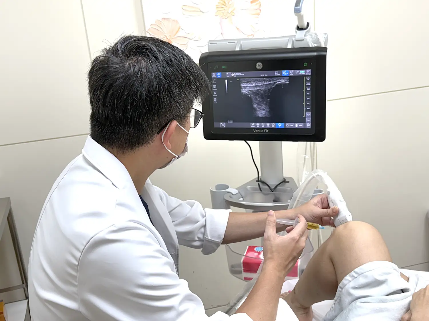
- Ultrasound-Guided Calcification Aspiration (Barbotage): Uses a needle to remove calcium deposits, often combined with PRP/PRF or high-concentration glucose to repair tendons, providing fast and significant results. (Learn more)
- Arthroscopic Shoulder Surgery: Removes calcium deposits and repairs tendons for patients with extensive deposits, recurrent flare-ups, or tendon tears.
Recovery and Prevention Tips:
- Post-surgery or post-removal treatment should be paired with rehabilitation to prevent adhesion and joint stiffness.
- Strengthening shoulder stabilizers (e.g., serratus anterior, scapular muscles) helps reduce recurrence.
- Avoid excessive overhead movements in daily life to reduce shoulder joint stress.
Early Diagnosis, Proper Treatment
Calcific tendinitis is one of the common causes of chronic shoulder pain and limited mobility. With early diagnosis and a variety of treatment options (such as shockwave/magnetic wave therapy, PRP/PRF, and ultrasound-guided aspiration), most patients can effectively relieve pain and restore their quality of daily life.
If you are experiencing shoulder pain, it is recommended to consult an orthopedic specialist to identify the true cause and receive appropriate treatment.
Frequently Asked Questions (FAQ) About Calcific Tendinitis
Q1: What is Calcific Tendinitis? Is it really like having \\\\\\"stones\\\\\\" in the shoulder?
Yes, it can be understood as \\\\\\"calcium crystals deposited within the tendon.\\\\\\" It most commonly occurs in the rotator cuff (especially the supraspinatus). The calcification causes friction and local inflammation, leading to pain, limited mobility, and in severe cases, affecting sleep and daily activities.
Q2: Does Calcific Tendinitis always cause pain? What are the typical symptoms?
Symptoms vary depending on the stage of calcification. Common presentations include:
- Sudden severe shoulder pain (even at rest)
- Difficulty raising the arm or putting on clothes
- Pain at night affecting sleep
Some people may feel almost nothing at the early stage, then experience sudden intense pain during the resorption phase.
Q3: Why did I think it was frozen shoulder, but it might be Calcific Tendinitis?
The symptoms are very similar: both can involve night pain, limited mobility, and difficulty raising the arm.
The differences are:
- Frozen Shoulder: Joint capsule adhesion and stiffness, usually \\\\\\"less painful at rest,\\\\\\" and others cannot help raise your arm much.
- Calcific Tendinitis: Tendon inflammation with calcification, often \\\\\\"painful even at rest,\\\\\\" sudden severe pain at certain angles, and others can usually help raise the arm a little.
Q4: Who is more prone to Calcific Tendinitis?
Common high-risk groups include:
- Women aged 30–50
- Throwing athletes (baseball, badminton, etc.)
- People who frequently lift heavy objects or perform overhead work (teachers, painters)
- Long-term keyboard users or those with tight shoulder/neck muscles
- People with a history of shoulder injury or recurrent inflammation
Q5: How is it diagnosed? Do I have to get an MRI?
Most cases do not require MRI. Common examinations include:
- X-ray: The most straightforward, often shows white calcification spots (like small stones)
- Ultrasound: Precisely locates the calcification, assesses tendon inflammation or tears, and can be used for guided treatment simultaneously
- MRI: Usually arranged only if tendon tear or other structural problems are suspected
Q6: Does Calcific Tendinitis always require surgery?
Not necessarily. Most patients can improve with non-invasive or minimally invasive treatments, such as:
- Shockwave Therapy (ESWT)
- Super Magnetic Wave Therapy (SIS)
- Rehabilitation and anti-inflammatory medications
Ultrasound-guided injection / calcification aspiration
Only a few cases with \\\\\\"large calcification, recurrent attacks, or tendon tears\\\\\\" may consider arthroscopic surgery.
Q7: What is Calcification Aspiration (Barbotage)? Is it fast-acting?
Barbotage uses a fine needle under ultrasound guidance to aspirate or flush out calcified deposits. It can be combined with PRP/PRF or prolotherapy to help tendon repair.
It is usually more effective when the calcification is in the resorption phase and softer, with many patients experiencing significant pain relief in a short period.
Q8: Can Calcific Tendinitis recur after treatment? How to prevent it?
Recurrence is possible, especially for those with long-term overuse, poor posture, or muscle imbalance. Recommendations:
- Follow rehabilitation after treatment to prevent adhesion and joint stiffness
- Strengthen shoulder stabilizers (serratus anterior, scapular muscles)
- Reduce prolonged overhead movements and repetitive friction
Doing these correctly can significantly reduce recurrence.
Related Articles
Please enter a title here Hsu Fang-wei (Orthopedic Surgeon at Jianli Clinic) | Orthopedic Specialist License No. 002188 Please enter a title here What Is Piriformis Syndrome? A Common Nerve Compression Problem in People Who Sit for Long Periods Every time you walk into an office or a coffee shop, t
Please enter a title here Hsu Fang-wei (Orthopedic Surgeon at Jianli Clinic) | Orthopedic Specialist License No. 002188 Please enter a title here Lately, in my clinic, I often see middle-aged women rush in, looking panicked, pointing to the middle joint of their finger and asking, “Doctor, is my fin
For degenerative knee osteoarthritis, conventional treatments can only relieve pain, whereas PRP (platelet-rich plasma) and PRF (platelet-rich fibrin) work through six scientifically proven mechanisms—“promoting cartilage regeneration,” “modulating inflammation,” and “improving mitochondrial functio


